
The future is digital
3D imaging, without the guesswork
Cone beam imaging (also called cone beam computed tomography, or CBCT) is one of the fastest-growing technologies in dentistry. It builds a detailed 3D digital model of your teeth, mouth, jaw and neck.
It is becoming the standard for planning dental implants, and it also helps with orthodontics, jaw joint (TMJ) problems, root canals, unusual teeth, and finding disease and cysts.
Up to 80% lower radiation
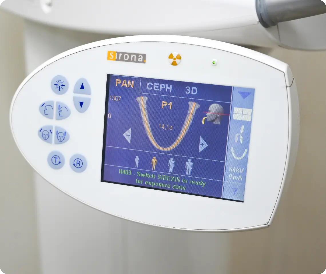
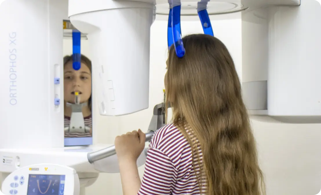
Images in about 15 seconds
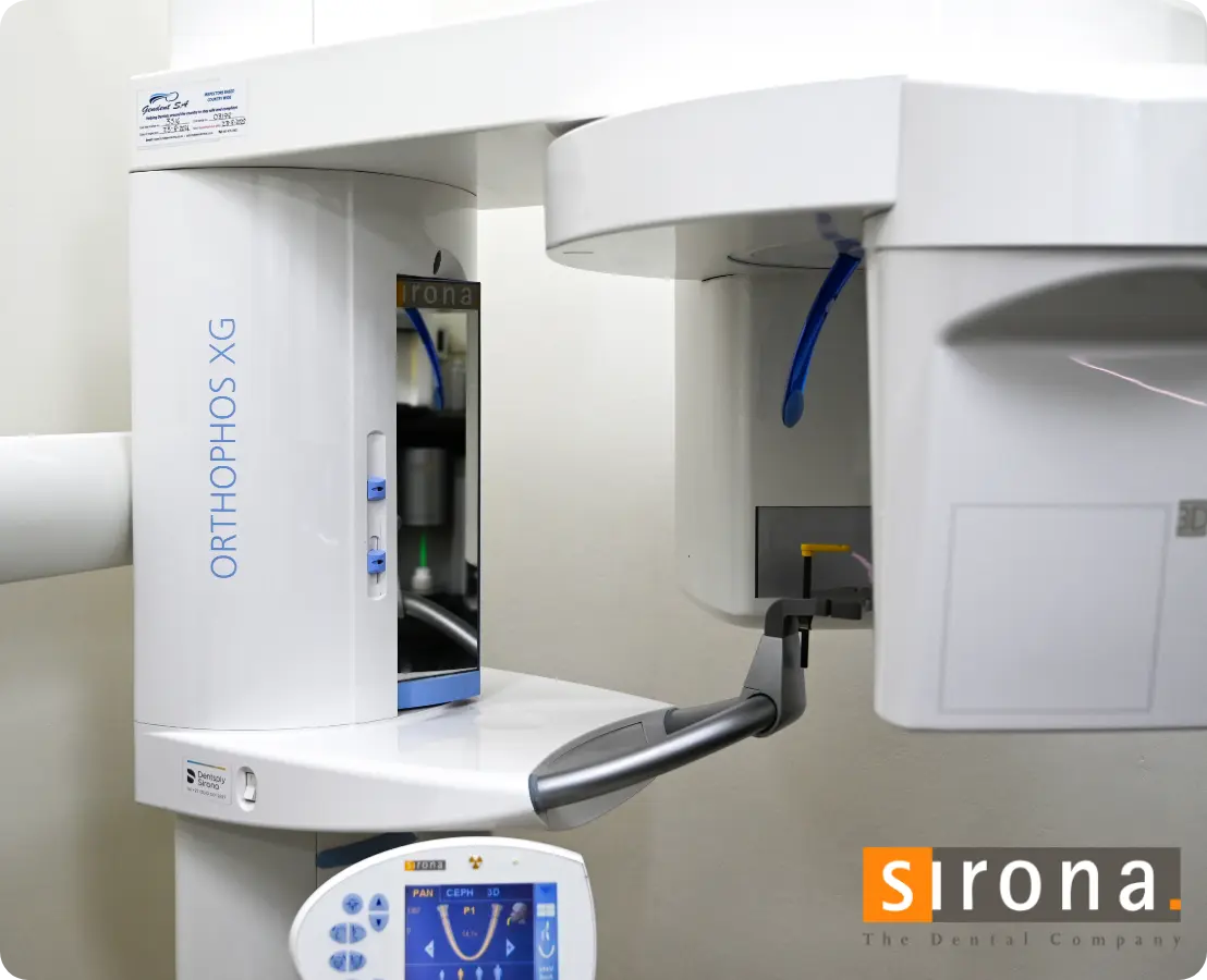
Open design, nothing closes in
Problems found earlier
How it helps
Every angle, on screen
Your dentist can bring up whichever view they need: 2D, 3D, a panoramic view of your whole mouth, even slices like the pages of a book. Images can be turned to any angle and magnified to see how bone, teeth, nerves and soft tissue relate, so treatment is planned and checked precisely.
More information
Radiation is a fair concern with any imaging. The newest cone beam systems use far lower doses and can focus on a small area, capturing only what’s needed.
Cone beam imaging grew out of Godfrey Hounsfield’s work on CAT scanning in the 1970s. A CAT scanner rotates a fan-shaped beam around the patient many times while moving them through the machine; a cone beam scanner rotates a cone-shaped beam and detector around you once, which is what makes it practical in a dental practice.
Book your appointment
Ready for your next check-up?
Book a visit at Alberton or Linksfield. Tell us what you need and the team will confirm a time with you.
